51+ Diagram Of Eye
Pads of fat and the surrounding bones of the skull protect them. Web Structure of the eye.

File Schematic Diagram Of The Human Eye With English Annotations Svg Wikipedia
The optic nerve is the largest sensory nerve of the eye.
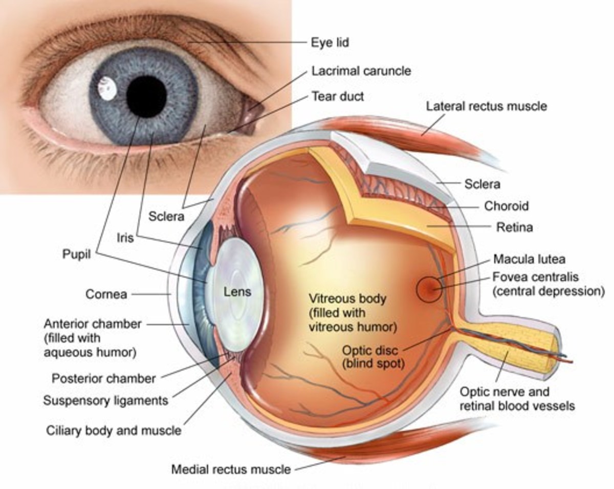
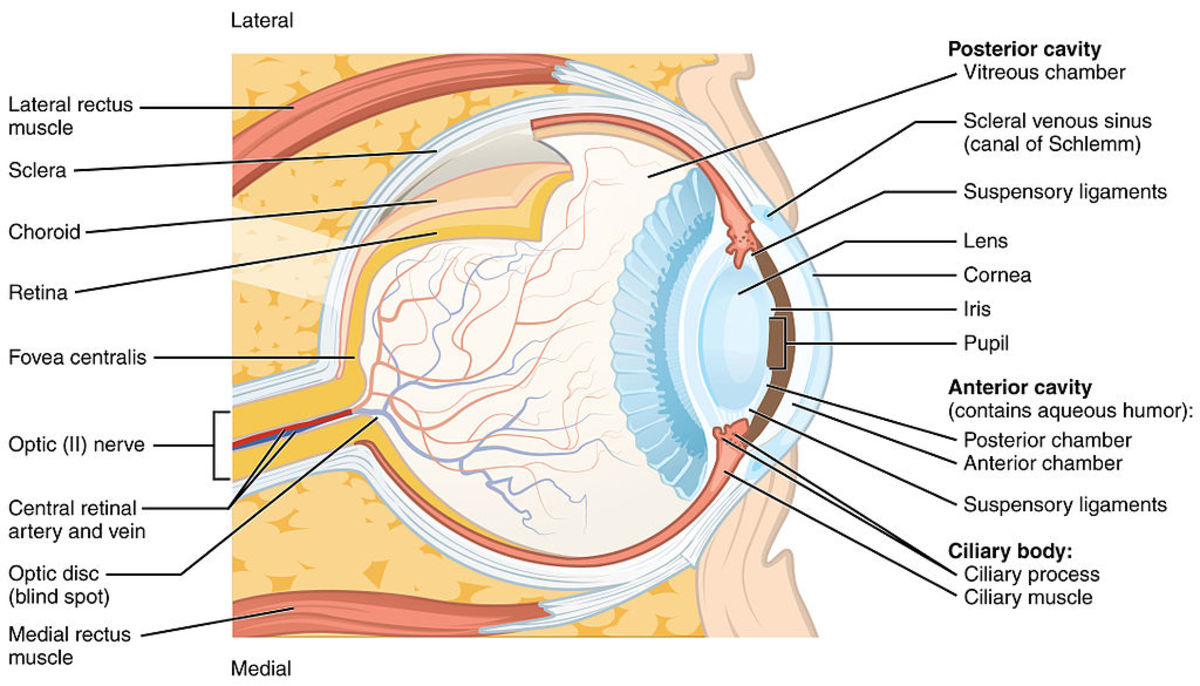
. The eye is a fluid-filled sphere enclosed by three layers of tissue Figure 111. It consists of the following parts. Web Diagram of a human eye horizontal section of the right eye 1.
Trabecular meshwork and Schlemms canal. The cornea pupil lens iris retina and. To understand the diseases and conditions that can affect the eye it helps to understand basic eye anatomy.
The optic nerve is the largest sensory nerve of. Muscles of the eye. Corneosclera or Fibrous tunic with 8.
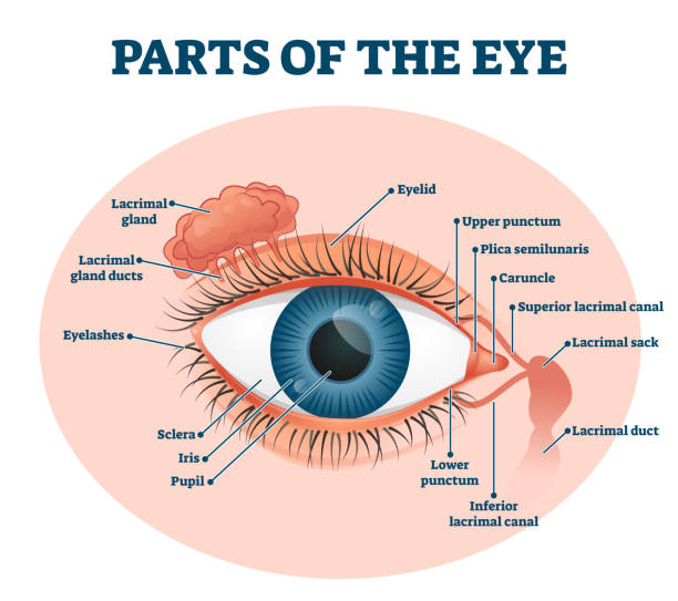
Check out this fact sheet to see a labeled diagram of the eye and learn about the different parts of the eye. Cornea Of The Eye. Web of light entering the eye.
The central aperture of iris is called pupil. The macula is the small sensitive area of the retina that gives central vision. Anterior chamber with 5.
It is located in the center of the retina. In higher organisms the eye is a complex optical system which. The anatomy of the eye includes auxiliary structures such as the bony eye socket and extraocular muscles as well as the structures of the eye itself such as the lens and the retina.
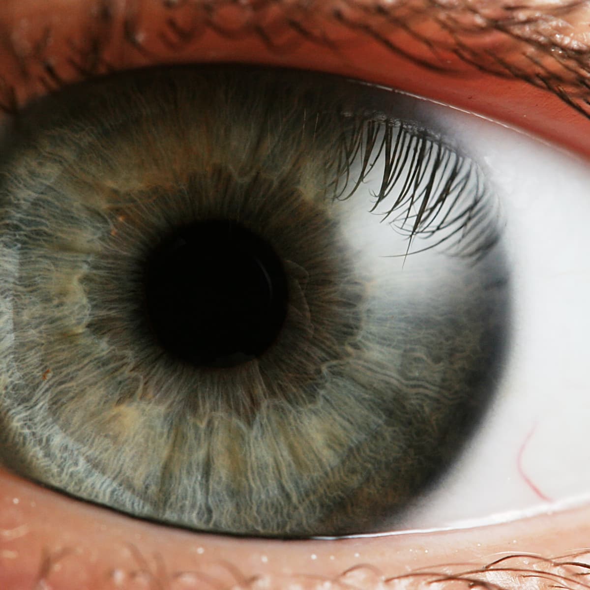
Corneal limbus and 11. The black circular opening in the iris that lets light in. Medial Rectus MR Moves the eye inward towards the nose.
Web amount of light entering the eye. The macula is the small sensitive area of the retina that. And for a description of common vision problems see Refraction and Refractive Errors.
When rays of light enter the eye theyre sort of. Here is a tour of the eye starting from the outside going in through the front and working to the back. It also contains a pigment that absorbs excess light so preventing blurring of vision.
The white of your eye. The White Of The Eye. Cornea of the eye.
The human eye is an organ that detects light and sends signals along the optic nerve to the brain. Cornea anterior chamber lens vitreous chamber and retina. Web Human eye specialized sense organ in humans that is capable of receiving visual images which are relayed to the brain.
It is composed of light sensitive cells known. The front transparent part of the sclera is called the cornea. A clear dome over the iris.
Innervation of the eye. The cornea has a curvature to it and covers the eye kind of like a crystal covering the face of a watch. The front section of the eyes interior where aqueous humor flows in and out providing nourishment to the eye.
The middle layer of the eye between the retina and the sclera. Web There are six extraocular muscles that attach to the outside of the eye from the bone in the eyes socket. Conjunctiva of the eye.
The clear watery fluid in the front of the eyeball. Schematic diagram showing a the orbital axis and the muscle axis at 51 for oblique muscles b with 51 adduction the orbital and muscle axis lie in the same plane and the muscle acts as a pure elevator depressor c with 39 abduction the orbital axis and muscle axis lie perpendicular to each other and muscle acts as a pure. It is the outer covering a protective tough white layer called the sclera white part of the eye.
It is located in the center of the retina. A human eye is roughly 23 cm in diameter and is almost a spherical ball filled with some fluid. Web This article explores the anatomy of the human eye looking at the different structures and their functions.
At the front of the eye however this opaque outer layer is transformed into the cornea a specialized transparent tissue that permits light rays to enter the eye. Web Eyes are organs of the visual systemThey provide living organisms with vision the ability to receive and process visual detail as well as enabling several photo response functions that are independent of visionEyes detect light and convert it into electro-chemical impulses in neurons neurones. Light reflects off the object were looking at and enters the eye through the cornea a clear thin dome-shaped tissue at the very front of the eye.
Perhaps one of the most complex organs of the body the eye is made up of several partsand each individual part contributes to your ability to see. These muscles work to rotate the eye and move the eye up down and from side to side. The white of the eye.
The lens is a clear part of the eye behind the iris that helps. The part of the eye that connects the choroid to the iris. Aperture Of The Eye.
Starting from front to back of the eye the cornea is in charge of shaping the light as it comes into the eye and passes through the lens. Web Anatomy of the Eye. When they work together these muscles can move the eye in any direction.
Posterior chamber and 4. Superior rectus inferior rectus medial rectus lateral rectus superior oblique inferior oblique levator palpebrae superioris Intrinsic. Web Anatomy of the Eye.
Most of the outer layer is composed of a tough white fibrous tissue the sclera. The diagrams show cross sections of the human eyeball. To focus light or an image on the retina.
Web View All. The eye has several major components. The Uvea Of The Eye.
A light sensitive layer that lines the interior of the eye. Web Eye color black brown blue etc is defined by the pigmentation of iris. Conjunctiva Of The Eye.
A thin layer of tissue. Eyes are approximately one inch in diameter. Sphincter pupillae dilator pupillae ciliaris.
Web Lauren Shavell Getty Images AARP. Zonule of Zinn or Ciliary zonule 3. It is circular in shape and allows light to pass through onto the lens.
Tubes arteries and veins that carry blood to and from the eye. Web Parts of the Eye. Web Major parts of the eye include the cornea pupil lens retina and macula.
The lens is a clear part of the eye behind the iris that helps to focus light or an image on the retina. Web Structure of Human Eye. Web Human eye anatomy seen from above For more details about specific structures of the eye and how they function visit these pages.
Web For more details about specific structures of the eye and how they function visit these pages. Aperture of the eye. As we journey through the different parts refer to them to better understand their functions.
Uvea of the eye.

Real Consumer Insights Pollfish Survey Tools
Anatomy And Structure Of The Human Eye With Diagrams Owlcation

Natural Vision Improvement Pinhole Glasses Http Altered States Net Index2 Php Healing Pinhole Htm Human Eye Diagram Eye Facts Eye Anatomy

The Alliance Skibidi Toilet Wiki Fandom

Simple Eye Diagram Easy Eye Diagram Labeled Eye Diagram Human Eye Diagram Eye Anatomy Diagram Eyeball Diagram

Formation Mechanism And Structure Of A Guanine Uracil Dna Intrastrand Cross Link Chemical Research In Toxicology
Anatomy And Structure Of The Human Eye With Diagrams Owlcation
Sports Free Full Text High Intensity Functional Training Perceived Functional And Psychosocial Health Related Outcomes From Current Participants With Mobility Related Disabilities

Simple Eye Diagrams Easy Eye Diagram Labeled Eye Diagram Human Eye Diagram Eye Structure Simple Eye

The Aging Surveyor Population Drones Technology And State Policy

Real Consumer Insights Pollfish Survey Tools

Does Perceptions Of Organizational Prestige Mediate The Relationship Between Public Service Motivation Job Satisfaction And The Turnover Intentions Of Federal Employees Leonard Bright 2021

1 700 Human Eye Diagram Stock Photos Pictures Royalty Free Images Istock Eye Chart Eye Ball
Anatomy And Structure Of The Human Eye With Diagrams Owlcation

Mohamed Abd El Mohdy Posted On Linkedin

File Eye Diagram No Circles Border Svg Wikipedia

Marketing Intern Job Description